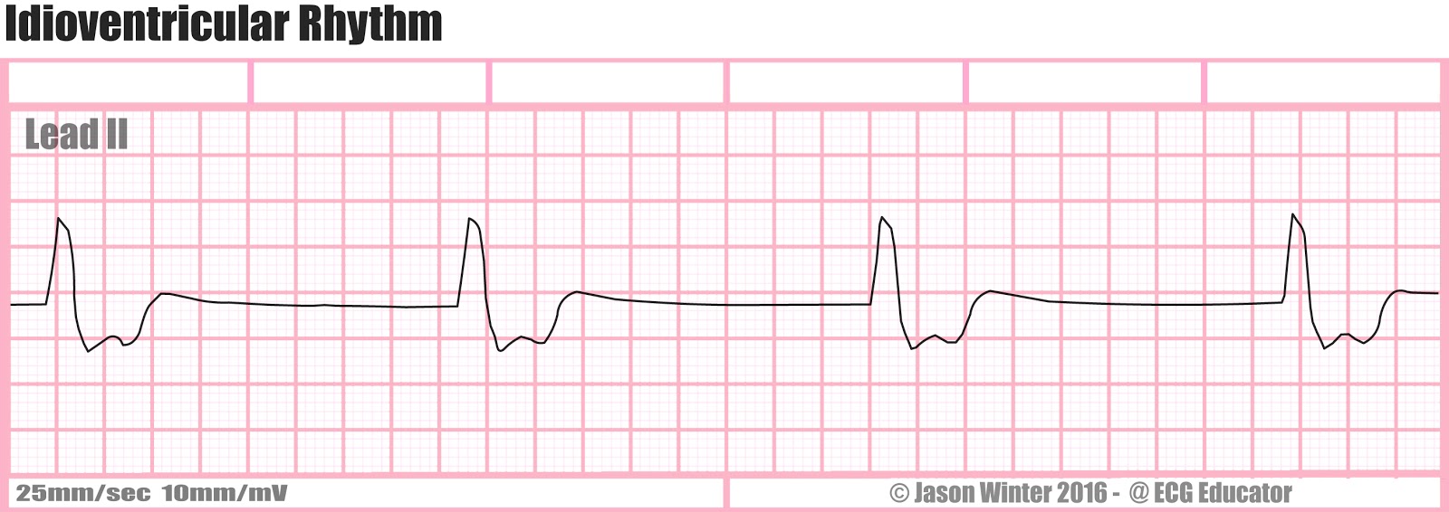
Idioventricular Rhythm
Accelerated Idioventricular Rhythm
Accelerated idioventricular rhythm is ventricular rhythm with a rate of between 40 and 120 beats per minute. Idioventricular means "“relating to or affecting the cardiac ventricle alone“ and refers to any ectopic ventricular arrythmia. Accelerated idioventricular arrhythmias are distinguished from ventricular rhythms with rates less than 40 (ventricular escape) and those faster than 120 (ventricular tachycardia). Though some other references limit to between 60 and 100 beats per minute. It is also referred to as AIVR and "slow ventricular tachycardia."
It can be present at birth. However, it is more commonly associated with reperfusion after myocardial injury.
Polymorphic Ventricular Tachycardia
Polymorphic ventricular tachycardia, on the other hand, has beat-to-beat variations in morphology. This may appear as a cyclical progressive change in cardiac axis, previously referred to by its French name torsades de pointes ("twisting of the spikes"). However, at the current time, the term torsades de pointes is reserved for polymorphic VT occurring in the context of a prolonged resting QT interval.
Ventricular Flutter
Ventricular Tachycardia (VT)
Monomorphic ventricular tachycardia means that the appearance of all the beats match each other in each lead of a surface electrocardiogram (ECG).Scar-related monomorphic ventricular tachycardia is the most common type and a frequent cause of death in patients having survived a heart attack or myocardial infarction, especially if they have weak heart muscle.
Ventricular Fibrillation (VF)


Ventricular fibrillation (V-fib or VF) is a condition in which there is uncoordinated contraction of thecardiac muscle of the ventricles in the heart, making them quiver rather than contract properly. Ventricular fibrillation is the most commonly identified arrhythmiain cardiac arrest patients. While there is some activity, the lay person is usually unable to detect it by palpating (feeling) the major pulse points of the carotid and femoral arteries. Such an arrhythmia is only confirmed by electrocardiography. Ventricular fibrillation is a medical emergency that requires prompt Advanced Life Support interventions. If this arrhythmia continues for more than a few seconds, it will likely degenerate further into asystole ("flatline"). This condition results in cardiogenic shock and cessation of effective blood circulation. As a consequence, sudden cardiac death (SCD) will result in a matter of minutes. If the patient is not revived after a sufficient period (within roughly 5 minutes at room temperature), the patient could sustain irreversible brain damage and possibly become brain-dead, due to the effects of cerebral hypoxia. On the other hand, death often occurs if sinus rhythm is not restored within 90 seconds of the onset of VF, especially if it has degenerated further into asystole.