Wednesday, 10 February 2016

Ventricular Rhythms


Idioventricular Rhythm

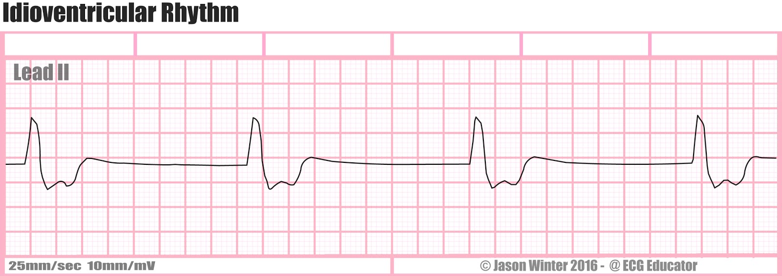

Accelerated Idioventricular Rhythm


Accelerated idioventricular rhythm is ventricular rhythm with a rate of between 40 and 120 beats per minute. Idioventricular means "“relating to or affecting the cardiac ventricle alone“ and refers to any ectopic ventricular arrythmia. Accelerated idioventricular arrhythmias are distinguished from ventricular rhythms with rates less than 40 (ventricular escape) and those faster than 120 (ventricular tachycardia). Though some other references limit to between 60 and 100 beats per minute. It is also referred to as AIVR and "slow ventricular tachycardia."


It can be present at birth. However, it is more commonly associated with reperfusion after myocardial injury.

Polymorphic Ventricular Tachycardia


Polymorphic ventricular tachycardia, on the other hand, has beat-to-beat variations in morphology. This may appear as a cyclical progressive change in cardiac axis, previously referred to by its French name torsades de pointes ("twisting of the spikes"). However, at the current time, the term torsades de pointes is reserved for polymorphic VT occurring in the context of a prolonged resting QT interval.


Ventricular Flutter


Ventricular Tachycardia (VT)

Monomorphic ventricular tachycardia means that the appearance of all the beats match each other in each lead of a surface electrocardiogram (ECG).Scar-related monomorphic ventricular tachycardia is the most common type and a frequent cause of death in patients having survived a heart attack or myocardial infarction, especially if they have weak heart muscle.


Ventricular Fibrillation (VF)


Ventricular fibrillation (V-fib or VF) is a condition in which there is uncoordinated contraction of thecardiac muscle of the ventricles in the heart, making them quiver rather than contract properly. Ventricular fibrillation is the most commonly identified arrhythmiain cardiac arrest patients. While there is some activity, the lay person is usually unable to detect it by palpating (feeling) the major pulse points of the carotid and femoral arteries. Such an arrhythmia is only confirmed by electrocardiography. Ventricular fibrillation is a medical emergency that requires prompt Advanced Life Support interventions. If this arrhythmia continues for more than a few seconds, it will likely degenerate further into asystole ("flatline"). This condition results in cardiogenic shock and cessation of effective blood circulation. As a consequence, sudden cardiac death (SCD) will result in a matter of minutes. If the patient is not revived after a sufficient period (within roughly 5 minutes at room temperature), the patient could sustain irreversible brain damage and possibly become brain-dead, due to the effects of cerebral hypoxia. On the other hand, death often occurs if sinus rhythm is not restored within 90 seconds of the onset of VF, especially if it has degenerated further into asystole.

Atrial Rhythms


Atrial Fibrillation (AF)


Atrial Flutter


Atrial Bigeminy


Supraventricular Tachycardia (SVT)


  1. Wandering Atrial Pacemaker (WAP)


Multifocal Atrial Tachycardia (MAT)


Premature Atrial Contraction (PAC)


Wolff-Parkinson White (WPW)


Junctional Rhythms


Junctional rhythm describes an abnormal heart rhythm resulting from impulses coming from a locus of tissue in the area of the atrioventricular node, the "junction" between atria and ventricles.

Under normal conditions, the heart's sinoatrial node determines the rate by which the organ beats - in other words, it is the heart's "pacemaker." The electrical activity of sinus rhythmoriginates in the sinoatrial node and depolarizes the atria. Current then passes from the atria through the bundle of His, from which it travels along Purkinje fibers to reach and depolarize the ventricles. This sinus rhythm is important because it ensures that the heart's atria reliably contract before the ventricles.

In junctional rhythm, however, the sinoatrial node does not control the heart's rhythm - this can happen in the case of a block in conduction somewhere along the pathway described above. When this happens, the heart's atrioventricular node takes over as the pacemaker. In the case of a junctional rhythm, the atria will actually still contract before the ventricles; however, this does not happen by the normal pathway of activation and instead is due to a backwards or retrograde conduction (conduction comes from the AV node into and through the atria).

Junctional rhythm can be diagnosed by looking at an ECG: it usually presents without a P wave or with an inverted P wave. Retrograde P waves refers to the depolarization from the AV node back towards the SA node.


Junctional Rhythm without P-waves

Junctional Rhythm with retrograde P-waves

Junctional Escape Rhythm

Accelerated Junctional Rhythm

Junctional Tachycardia

Premature Junctional Complex (PJC)


Sinoatrial Node rhythms

The sinus node (SA) is located in the roof of the right atrium. It is the fastest physiological pacemaker. When the sinus node generates an electrical impulse, the surrounding cells of the right atrium depolarize. Then the cells of the left atrium, the AV (atrioventricular) node, follow, and at last the ventricles are stimulated via the His bundle. 


Normal Sinus Rhythm (NSR)


The term normal sinus rhythm (NSR) is sometimes used to denote a specific type of sinus rhythm where all other measurements on the ECG also fall within designated normal limits, giving rise to the characteristic appearance of the ECG when the electrical conduction system of the heart is functioning normally.[2] However, other sinus rhythms can be entirely normal in particular patient groups and clinical contexts, so the term is sometimes considered a misnomer and its use is sometimes discouraged.


Sinus Bradycardia

Sinus bradycardia is a sinus rhythm with a rate that is lower than normal. In humans, bradycardia is generally defined to be a rate of under 60 beats per minute.

Sinus Tachycardia

Sinus tachycardia (also colloquially known as sinus tach or sinus tachy) is a sinus rhythm with an elevated rate of impulses, defined as a rate greater than 100 beats/min (bpm) in an average adult. The normal resting heart rate in the average adult ranges from 60–100 beats/min. Note that the normal heart rate varies with age, with infants having normal heart rate of 110–150 bpm, in contrast to the elderly, who have slower normals.

Sinus Arrhythmia

Respiratory sinus arrhythmia (RSA) is a naturally occurring variation in heart rate that occurs during a breathing cycle. RSA is also a measure of parasympathetic nervous system activity - which denotes "rest and digest" behaviors.


Sinus Exit Block


Sinus Pause